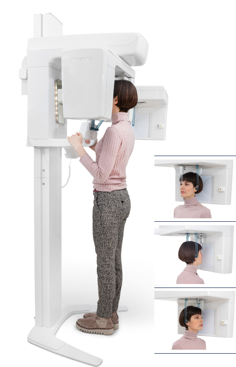
X-VIEW: Efficient and modern digital radiographic system
Efficient and modern digital radiographic system to provide doctors with an extremely useful tool for accurate diagnostics and successful treatments. OPG/CBCT technology combines panoramic and cone beam computed tomography (CBCT) imaging to provide comprehensive diagnostic capabilities. This dual-function system allows dental professionals to capture both 2D panoramic images and 3D CBCT scans, offering a more complete view of the patient’s oral structures. The detailed imaging enhances the ability to diagnose complex cases such as impacted teeth, jaw disorders, and airway assessments. With reduced radiation exposure and high image clarity, OPG/CBCT is ideal for a wide range of dental procedures. This technology is designed to streamline workflow and improve patient outcomes by delivering accurate and timely diagnoses.
HIGHLIGHTS
- CMOS Scanning detector, Complementary Metal Oxide Semiconductor (CMOS) sensor offers:
• Higher Sensitivity due to the latest pixel architecture which is beneficial in lower light applications
• Lower dark noise that contributes to a higher fidelity image
• Improved pixel well depth (saturation capacity) that provides higher dynamic range
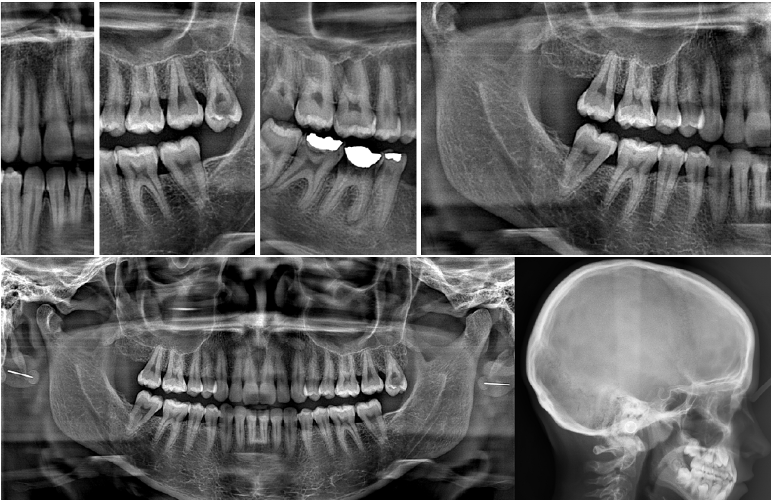
• Lower Power consumption (eco-green) - More comfort for patients since Panoramic X-rays are extraoral and simple to perform. Improved Care. A panoramic X-ray allows the view of the head, neck, and jaw, and how they work together as a whole, which means doctors can more easily:
• Assess patients with an extreme gag reflex
• Evaluate the progression of TMJ
• Expose cysts and abnormalities
• Expose impacted teeth
• Expose jawbone fractures
• Plan treatment (full and partial dentures, braces
and implants)
• Reveal gum disease and cavities - ADVANCED 2D AND CEPH SOFTWARE FUNCTIONS: Full connectivity to easily export, share and store images. Uses Deep- View software and the same database for both 2D and Ceph programs.
- DR CEPH DETECTOR ADVANTAGES
• Better contrast
• More details and filtering
• No background disturbance
• Exposure time: 200-500 ms
• Reading time: immediate
• Detector-PC image transmission: 2sec
• Calibration method: easy, intuitive and manageable remotely - Elegant and compact Italian design
QUICK AND EASY PATIENT POSITIONING
X-VIEW 2D PAN provides great stability to patients, quick access and a natural body position. Ergonomics in design helps to achieve the perfect focal trough and correct patient’s posture. The specially designed tools assist patients, during the acquisition process, to maintain their position
avoiding errors in the procedures:
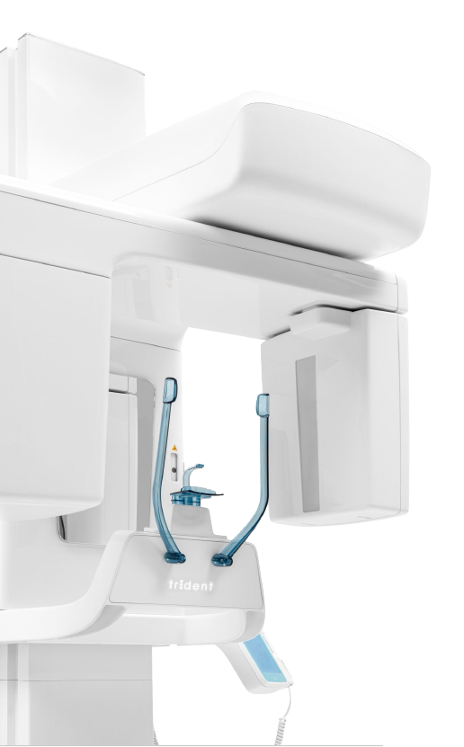
• The bite block centers the teeth by aligning the arches, positioning the incisors on the focal plane,
and allowing vertical symmetry
• Two laser lines trace the references of the area of interest, while the mirror located in front of the
bite-block helps patients to control their position
X-VIEW 2D PAN adapts to all sizes and types of patients, the linear and open design facilitates access for wheelchair users. The X-VIEW 2D PAN motorized lifting with two speeds makes patient positioning easier than ever. The quick movement allows adjustment of the unit to the patient height; with the slow movement a precise alignment is achieved using the laser. The double laser is an essential tool to get the perfect inclination and orientation of the patient’s head. The chin rest helps to achieve an accurate focal plane for very low risk of error in diagnostic images.
